
What is ARMD
Age-related macular degeneration (ARMD) is an acquired retinal degeneration that significantly impairs central vision as a result of both non-neovascular (abnormalities of the drusen and retinal pigment epithelium) and neovascular derangements (choroidal neovascular membrane formation). Subretinal fibrosis, bleeding beneath the retina or behind the retinal pigment epithelium (RPE), and serous fluid may all be signs of advanced illness. Although environmental, dietary, and developmental (i.e., aging) mechanisms combine to alter the degeneration seen in the macula, a genetic component is postulated given its propensity to people of European ancestry. There is a great need for innovative medicines due to recently implicated biochemical pathways and the lack of effective treatments for the majority of armds (i.e., dry ARMD).
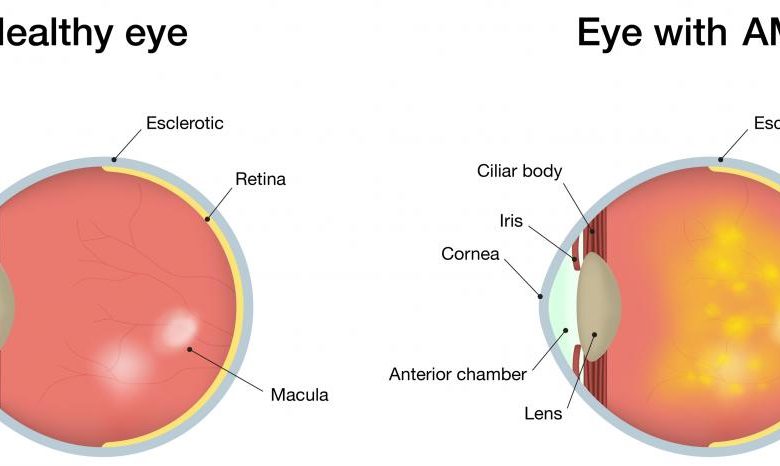
AMD Signs and Symptoms
Dry AMD
Most patients still have enough vision to read and drive despite the gradual, painless loss of central vision. Scotomas, or central blind patches, typically appear late in the course of the illness and can occasionally be severe. Typically, symptoms are bilateral.
The following alterations are caused by funduscopic changes:
- Retinal pigment epithelium changes
- Drusen
- Areas of chorioretinal atrophy
Wet AMD
Wet AMD is more common when vision loss occurs suddenly, typically spanning days to weeks. Visual distortion, such as a central blind spot (scotoma) or curving of straight lines, is frequently the initial symptom (metamorphopsia).
Color and peripheral vision are typically unaffected; nevertheless, if AMD is left untreated, the patient runs the risk of become legally blind (vision of 20/200 or less) in the affected eye. Wet AMD symptoms are frequently unilateral since it typically only affects one eye at a time.
The following alterations are caused by funduscopic changes:
- Localized retinal elevation caused by subretinal fluid
- Retinal swelling
- Discoloration under the macula that is gray-green
- Macula exudates or exudates nearby
- Retinal pigment epithelium separation (visible as an area of retinal elevation)
- Subretinal bleeding in or near the macula.
Diagnosis & Tests
When you see our best eye doctor (at bharti eye foundation) for a standard eye exam and have your eyes dilated, they can screen you for age-related macular degeneration. An early diagnosis will allow you to begin therapy, which could delay certain symptoms or lessen their severity.
Tests
Your retina—a layer of tissue at the back of your eye that processes light—will be examined, along with your eyesight and eye pressure. Under the retina, they’ll search for drusen, which are small yellow deposits. It’s a typical early indication of the sickness. Your physician might also ask you to examine an Amsler grid, which is a checkerboard-like pattern made up of straight lines.
Macular degeneration may be indicated if you notice that some of the lines are absent or appear to be wavy.
Tests If our doctor suspects you have age-related macular degeneration, they might recommend one or both of the following tests:
- Optical coherence tomography (OCT). A magnified 3D image of your retina may be seen in this unique photograph. Using this technique, your doctor can determine whether the retinal layers are misaligned. Additionally, whether you underwent laser or injectable therapy, they can determine whether the edema is becoming better or worse.
- Imaging with fluorescein. In this treatment, a dye is injected into a vein in your arm by your doctor.
They capture images as the dye enters your eye and travels through the retina’s blood vessels. Images of the macula, a small region near the center of your retina, will reveal new veins or vessels that are leaking blood or fluid.
Prevention
When you visit your doctor for an annual eye exam and a dilated exam, they can examine your eyes for age-related macular degeneration.
An early diagnosis will allow you to begin therapy, which could delay certain symptoms or lessen their severity.
In addition to testing your eyesight, they’ll look at your retina, a layer of tissue at the back of your eye that deals with light. Under the retina, they’ll search for drusen, which are small yellow deposits.
It’s a typical early indication of the sickness. Your physician might also ask you to examine an Amsler grid, which is a checkerboard-like pattern made up of straight lines. Macular degeneration may be indicated if you notice that some of the lines are absent or appear to be wavy.
Exams If our best eye doctor suspects you have age-related macular degeneration, they might recommend one or both of the following tests:
- With optical coherence tomography (OCT). A magnified 3D image of your retina may be seen in this unique photograph. Using this technique, your doctor can determine whether the retinal layers are misaligned. Additionally, whether you underwent laser or injectable therapy, they can determine whether the edema is becoming better or worse.
- Fluorescein angiography. In this treatment, a dye is injected into a vein in your arm by your doctor. They capture images as the dye enters your eye and travels through the retina’s blood vessels. Images of the macula, a sliver of the retina at its center, will reveal new arteries or vessels that are dripping blood or fluid
Read More: